New Patients
(865) 876-7050
Existing Patients
(865) 947-2220

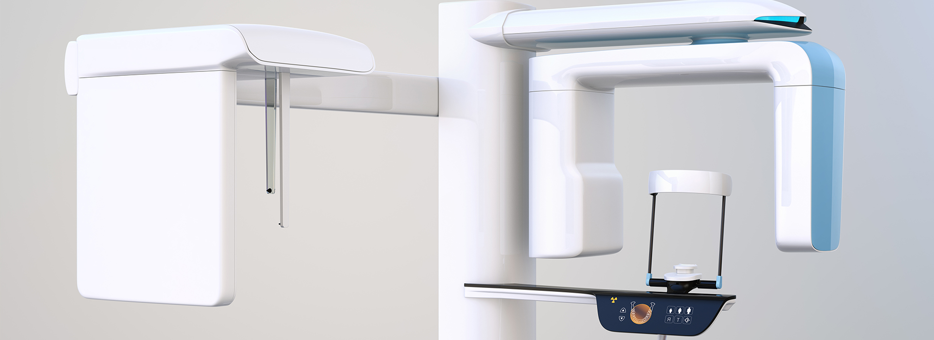
At Kennedy Dentistry, we combine clinical experience with advanced imaging to provide precise, patient-focused care. Cone-beam computed tomography (CBCT) gives our clinicians a three-dimensional view of teeth, jawbone, nerves, and sinuses — information that simply isn’t available from conventional two-dimensional X-rays. Having access to high-resolution 3D scans helps our team make confident diagnoses and plan treatments with greater predictability.
Our practice uses a modern CBCT system that balances clear, diagnostic-quality images with thoughtful attention to patient safety and comfort. The following sections explain how CBCT works, when it is most useful, and what patients can expect during a scan. The goal is to demystify the technology so you can feel informed and comfortable about its role in your care.
CBCT captures volumetric data in a single, rotating scan, creating a detailed 3D model of the head and neck region. This model shows the exact position and shape of teeth, the density and contour of the jawbone, the course of nerve canals, and nearby sinus spaces. Because the images are distortion-free and spatially accurate, clinicians can measure distances, angles, and volumes with confidence.
These scans make it easier to detect conditions that might be missed or underappreciated on traditional X-rays, such as root fractures, impacted teeth in complex positions, or small areas of bony loss. For patients who have had previous dental work, CBCT can clarify the relationship between restorations, implants, and surrounding bone, enabling safer, more informed decision-making.
Importantly, CBCT images are cross-sectional and can be viewed in multiple planes (axial, coronal, sagittal) or reconstructed into realistic 3D renderings. That flexibility helps both clinicians and patients visualize anatomy and pathology more readily than flat images, which improves communication and reduces uncertainty about recommended treatments.
One of CBCT’s most transformative uses is in planning implant placement and other oral surgeries. The scan provides exact measurements of bone height, width, and density, which are essential for choosing implant size and angulation and for identifying areas where bone grafting may be necessary. Planning with CBCT reduces surprises during surgery and supports more predictable, long-term outcomes.
Beyond measurements, CBCT helps map the location of critical anatomical structures, such as the inferior alveolar nerve and maxillary sinuses. Knowing these relationships ahead of time allows the surgical team to design an approach that minimizes risk while maximizing functional and esthetic results. In complex cases, CBCT data can be used to fabricate surgical guides that translate virtual plans precisely into the clinical setting.
Since CBCT delivers a comprehensive view of the operative field, it also aids in staging multidisciplinary care. For patients who require combined periodontal, restorative, and surgical procedures, the 3D data streamline coordination among specialists and help ensure that each phase of treatment aligns with the overall plan.
Contemporary CBCT systems are engineered to capture focused regions at the lowest dose necessary for the diagnostic task. Unlike traditional medical CT scanners that use higher radiation for large fields of view, dental CBCT units are optimized for targeted imaging. This means clinicians can choose smaller fields and tailored settings to limit exposure while still obtaining diagnostically useful images.
Clinical protocols emphasize justification and optimization: CBCT is used when the additional information it provides will directly influence diagnosis or treatment. For routine exams where two-dimensional films suffice, CBCT is not routinely indicated. When a 3D scan is recommended, the team will explain the reasons and how the chosen scan parameters protect patient safety.
Alongside exposure considerations, CBCT appointments are efficient. Scans typically take only a few seconds of active imaging time, and patients remain stationary while the machine rotates. The short duration reduces motion artifacts, improving image clarity and reducing the need for repeat exposures.
CBCT is versatile and supports decision-making across many areas of dentistry. Endodontists use it to locate hidden canals, assess periapical pathology, and evaluate complex root anatomy. Oral surgeons rely on it for impacted tooth extractions, pathology assessment, and trauma evaluation. Orthodontists and restorative dentists use 3D data to evaluate tooth position, airway relationships, and the skeletal framework underlying the smile.
The ability to inspect bone volume and quality also makes CBCT valuable in periodontal assessment and in monitoring healing after grafting or implant placement. When pathology is suspected, such as cysts or unusual bone changes, three-dimensional imaging gives a clearer picture of extent and relation to adjacent structures than flat radiographs can provide.
Because CBCT data are digital and interoperable, images can be reviewed collaboratively with specialists when needed. Sharing standardized 3D datasets enhances multidisciplinary planning and ensures that all members of a treatment team are working from the same precise information.
From a patient perspective, CBCT appointments are straightforward: you sit or stand while the scanner rotates around your head, and there is no need for claustrophobic enclosures or lengthy procedures. The noninvasive nature of the scan, combined with its speed, often makes it a comfortable option even for patients with dental anxiety.
Once images are captured, clinicians can use clear visual aids — sliced views and rendered 3D models — to explain findings and outline treatment options. Visualizing anatomy in three dimensions helps patients understand the rationale for recommended care and the expected steps involved in their treatment plan. This clarity supports more informed consent and fosters collaboration between patient and provider.
Our approach emphasizes education and transparency. If CBCT is recommended, the team will review the images with you, describe what is being evaluated, and discuss how the information will be used to improve your care. That open dialogue helps patients feel confident in the proposed path forward.
In summary, CBCT is a powerful diagnostic tool that enhances accuracy, improves surgical planning, and supports safer, more predictable dental care. When used thoughtfully and selectively, three-dimensional imaging provides clinicians and patients with a clearer understanding of anatomy and treatment options. If you’d like to learn more about how CBCT might be used in your care, please contact us for more information.
